Image-guided therapy
Interventional imaging systems, smart devices, software and services
Image-guided therapy
Interventional imaging systems, smart devices, software and services
Image-guided therapy
Interventional imaging systems, smart devices, software and services
Image-guided therapy portfolio
See clearly. Treat optimally.
Every day you navigate some of life’s most challenging situations amidst the complexities of modern healthcare. Your work inspires us to develop more seamless solutions – to help you decide, guide, treat, and then confirm the right care, for every patient, in real time.
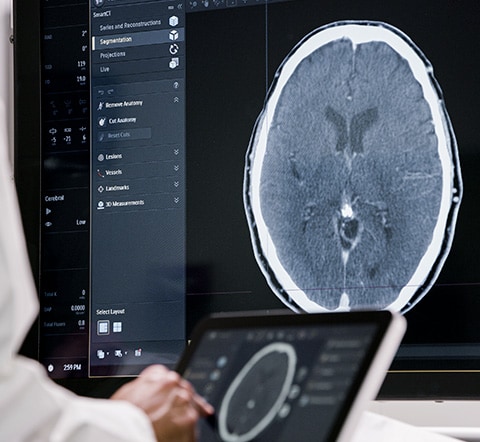
Today, the Philips portfolio of image-guided therapy (IGT) solutions uniquely integrates best in class imaging systems and software, with specialized diagnostic and therapeutic devices to support exceptional treatment for even the most complex procedures.
What’s next? More meaningful innovation for more interventional procedures, and continuing support for you with evidence-based technologies to simplify workflows, enhance patient care and reduce costs.
Together, we make life better.
Hear from key opinion leaders

Dr. Carlos E. Ruiz
MD, PhD, Chair, Structural Heart and Congenital Heart Disease, Hackensack University Medical Center
Customer perspectives
Physicians share the ways Philips image-guided therapy innovations deliver value in the diagnosis and treatment of patients.
Prof. Spelle
Interventional Neuroradiologist, Chairman NEURI, the Brain Vascular Centre Hôpital Bicêtre AP-HP, Paris France
Perspectives in image-guided therapy
Explore our customer success stories and insightful articles from leaders in image-guided therapy
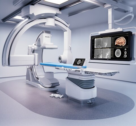
Innovatively designed to support even the most challenging procedures
Working in close collaboration with healthcare professionals, we keep enhancing the Philips Azurion experience so you can perform a wide range of routine and complex interventional procedures easily and confidently, now and in the future.
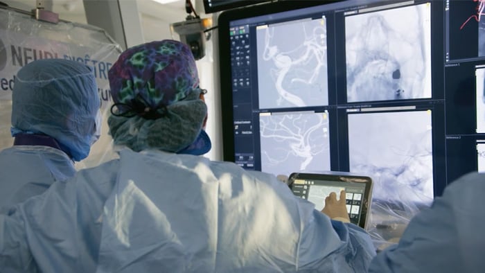
Faster stroke treatment. Learn more about Direct to Angio Suite workflow.
When it comes to acute stroke, everyone deserves the best care. Today, more patients can benefit from mechanical thrombectomy through changed guidelines*, which widen the time window for endovascular treatment.
Extensions to our image-guided therapy portfolio
Supporting you at every turn
DoseWise
IGT upgrades
IGT services
IGT refurbished systems
Philips strategic partnerships
IGT Systems education
IGT Devices education
News and blogs on image-guided therapy


* View guidelines here: www.philips.com/dtas