- High resolution EEG data
-
High resolution EEG data
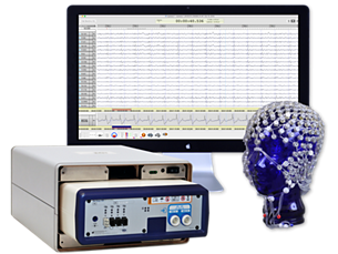
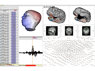
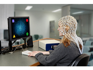
To provide the best source imaging results, GeoSource 3 Research software makes full use of high density EEG data, with up to 256 sensors and whole head coverage. - Highly realistic head anatomy
-
Highly realistic head anatomy
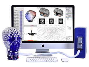
Faithful head anatomy is critical for accurate source imaging. Automated software characterizes tissue segmentation — scalp, skull, cerebrospinal fluid, grey matter, white matter, air, and eyeballs — directly from 1 mm MRI data for the most realistic anatomy across the entire head. Head model can be built from individual MRI data. - Detailed current flow models
-
Detailed current flow models
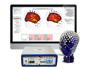
A high-resolution model of the current flow through the head is key to accurate source imaging results. The Finite Difference Method (FDM) provides voxel-by-voxel calculation of electrical potentials, maintaining the high resolution geometry of the original MRI image. - Individual head model building
-
Individual head model building
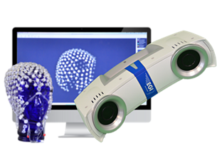
Dedicated pipeline software guides you through each step to build an individual FDM head model with opportunities to review and edits the model at all stages of construction. Compute time is accelerated from days to minutes using the included high-performance GPU. Obtain more precise data with 3D sensor positions to match the patient’s head geometry. - 6 atlas head models
-
6 atlas head models
Even without an individual MRI scan, you can use the 6 built-in age and gender specific atlas head models to get faster source imaging results. - Your choice of three different packages
-
Your choice of three different packages
6 built-in age and gender specific atlas head models in the BASIC package. Use digitized sensor positions to conform/warp an atlas head model to individual head geometry (requires sensor registration) with the INTERMEDIATE package. Import MRI scans for the most realistic brain anatomy to build individual head models with the ADVANCED Package.
High resolution EEG data
High resolution EEG data
Highly realistic head anatomy
Highly realistic head anatomy
Detailed current flow models
Detailed current flow models
Individual head model building
Individual head model building
6 atlas head models
6 atlas head models
Your choice of three different packages
Your choice of three different packages
- High resolution EEG data
- Highly realistic head anatomy
- Detailed current flow models
- Individual head model building
- High resolution EEG data
-
High resolution EEG data
To provide the best source imaging results, GeoSource 3 Research software makes full use of high density EEG data, with up to 256 sensors and whole head coverage. - Highly realistic head anatomy
-
Highly realistic head anatomy
Faithful head anatomy is critical for accurate source imaging. Automated software characterizes tissue segmentation — scalp, skull, cerebrospinal fluid, grey matter, white matter, air, and eyeballs — directly from 1 mm MRI data for the most realistic anatomy across the entire head. Head model can be built from individual MRI data. - Detailed current flow models
-
Detailed current flow models
A high-resolution model of the current flow through the head is key to accurate source imaging results. The Finite Difference Method (FDM) provides voxel-by-voxel calculation of electrical potentials, maintaining the high resolution geometry of the original MRI image. - Individual head model building
-
Individual head model building
Dedicated pipeline software guides you through each step to build an individual FDM head model with opportunities to review and edits the model at all stages of construction. Compute time is accelerated from days to minutes using the included high-performance GPU. Obtain more precise data with 3D sensor positions to match the patient’s head geometry. - 6 atlas head models
-
6 atlas head models
Even without an individual MRI scan, you can use the 6 built-in age and gender specific atlas head models to get faster source imaging results. - Your choice of three different packages
-
Your choice of three different packages
6 built-in age and gender specific atlas head models in the BASIC package. Use digitized sensor positions to conform/warp an atlas head model to individual head geometry (requires sensor registration) with the INTERMEDIATE package. Import MRI scans for the most realistic brain anatomy to build individual head models with the ADVANCED Package.
High resolution EEG data
High resolution EEG data
Highly realistic head anatomy
Highly realistic head anatomy
Detailed current flow models
Detailed current flow models
Individual head model building
Individual head model building
6 atlas head models
6 atlas head models
Your choice of three different packages
Your choice of three different packages
Documentation
-
Brochure (1)
-
Brochure
- GeoSource 3 Research (1.2 MB)
-
User manual (1)
-
User manual
- GeoSource 3 Research (19.6 MB)
-
Brochure (1)
-
Brochure
- GeoSource 3 Research (1.2 MB)
-
User manual (1)
-
User manual
- GeoSource 3 Research (19.6 MB)
-
Brochure (1)
-
Brochure
- GeoSource 3 Research (1.2 MB)
-
User manual (1)
-
User manual
- GeoSource 3 Research (19.6 MB)
Related products
Alternative products
-
Geodesic EEG System 400 Research MR conditional kit
- One system for routine EEG or simultaneous EEG-fMRI
- High density EEG systems compatiable with fMRI
- Use special MR compatible EEG sensors, amplifier, and software
- MR compatible saline or gel Nets products
View product
-
Geodesic EEG System 400 Research
- • Whole head, high density EEG system for advanced brain research
- • Easy-to-apply sensors, intuitive software designed for review and analysis of high density EEG
- • Supports EEG-fMRI, EEG-MEG, EEG-TES and EEG-TMS
- • Interoperability with EEGLAB
View product
-
GTEN 100 Neuromodulation Research System
- Engineered for precise, reproducible, personalized neuromodulation
- True HD TES with up to 256 electrodes
- Uses the same electrode net for tDCS, tACS, tPCS, tRNS and simultaneous EEG
- Designed to maximize current to the target while minimizing current elsewhere
View product
-
GeoScan Research
- Real time 3D sensor map creation for up to 256 EEG sensors
- Minimal time for both participant and operator
- High accuracy
- Compact size and mobility
View product
-
GeoSource 3 Research ESI software
- High density ESI software for 256 channels of EEG
- Uses highly realistic head models created from 1-mm MRI data, 7 characterized tissue types and FDM
- Choice of individual, age and gender specific atlases, or conformal atlas head models
View product
-
Geodesic Photogrammetry System 3 Research (GPS)
- Instantaneous imaging for up to 256 EEG sensors
- Minimal participant time
- Semi-automated 3D sensor map creation
- High accuracy
View product
-
E-Prime
- Software and hardware for stimulus presentation experiments
- Fully integrated into Net Station
- Chronos: Powerful new USB-based response pad and auditory stimulus presentation
View product
-
Geodesic EEG System 400 Research MR conditional kit
The ideal platform for high density EEG-fMRI. MR conditional EEG sensors, amplifier, and software work together to minimize and remove imaging-related artifacts.
View product
-
Geodesic EEG System 400 Research
The advanced Geodesic EEG System (GES) makes whole head, high density EEG accessible for any research lab, with fast-to-apply and comfortable sensor nets plus intuitive software. The expandable, modular product design and compatibility with open-source software tools provides a flexible platform that can grow with your lab and research needs.
View product
-
GTEN 100 Neuromodulation Research System
With the GTEN (Geodesic Transcranial Electrical Neuromodulation) 100 neuromodulation research system, achieve true high-definition electrical neuromodulation. Use the same HD EEG Geodesic Sensor Nets to record EEG signals, localize cortical activity, and stimulate brain regions of interest with a variety of protocols.
View product
See all related products -
GeoScan Research
Real time 3D sensor map creation. Creates a 3D coordinate file of up to 256 sensor locations in ~15 minutes.
View product
-
GeoSource 3 Research ESI software
GeoSource 3 Research software leverages high density EEG technology, high-resolution MRI imaging, sophisticated electric head modeling, and accelerated computing to create a powerful platform for electrical source imaging, support for advanced neuromodulation planning and other advanced research applications.
View product
-
Geodesic Photogrammetry System 3 Research (GPS)
Simultaneously record the location of all sensors on the head with the click of a button. Solve for sensor locations at any time from the saved photographs. Designed to improve source imaging and minimize participant time.
View product
-
E-Prime
A suite of applications that allows you to design, generate, and run behavioral and ERP experiments with seamless, millisecond precision, integration with Net Station research software The graphical interface makes it straightforward to build your own experiments, deliver stimuli, collect responses, and edit and analyze data.
View product
- This instrument is not intended for use in diagnosis or treatment of any disease or condition. It is a scientific research instrument designed for performing measurements and acquiring data for neurophysiological research. Philips makes no representation of the suitability of the instrument for any particular research study.
